
Figure 3.
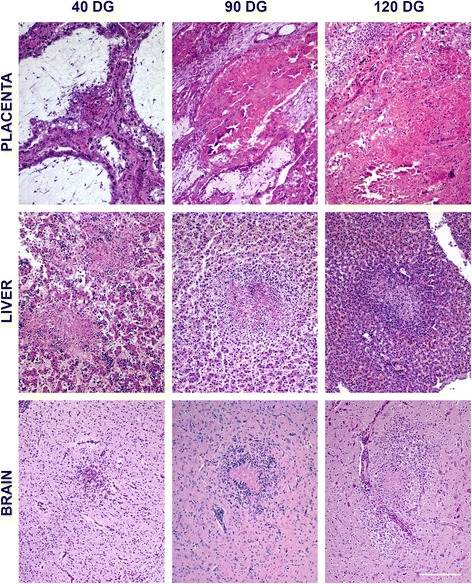
Comparison of the characteristic microscopic lesions found in placenta and foetal liver and brain. Pictures show the histological changes measured for the quantification of lesions. When necrotic areas and infiltration of inflammatory cells coexisted, mainly at the liver and brain of G2 and G3, only the area showing necrosis was measured. All the pictures were taken at the same magnification. Bar 200 μm.