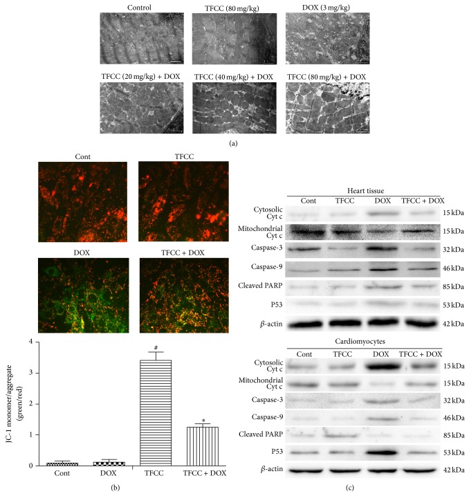
Figure 7.
Effects of DOX and TFCC on mitochondrial apoptotic pathway in vivo and in vitro. Rats were treated with vehicle or DOX (i.p., 3 mg/kg every other day) with or without TFCC pretreatment (20, 40, and 80 mg/kg i.p.). At day 14, the ultrastructure of cardiomyocytes was observed using electron microscope (a). H9c2 cells were treated with vehicle or DOX (1 μM) with or without TFCC (25 μg/mL for 4 h prior to DOX exposure). After 24 h, cells were stained with JC-1 dye and were visualized by fluorescence microscopy. Quantitative analysis of JC-1 staining was evaluated (green to red fluorescence ratio) (b). Cytochrome c (cyt c) in mitochondrial and cytosolic protein extracts and caspase-3/9 and PARP levels in rat heart tissues and H9c2 cells were determined using Western blot analysis (c). Cont, vehicle treatment; TFCC, TFCC treatment; DOX, DOX treatment; TFCC + DOX, TFCC and DOX cotreatment. The results are represented as the mean ± SE. # P < 0.05 relative to the control group, and * P < 0.05 relative to the DOX group.

