Figure 3.
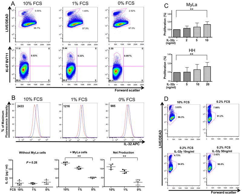
IL-32 facilitates cell proliferation and augments viability of CTCL cells. A, After MyLa cells were cultured in media with the indicated FCS concentrations for 40 hours, they were stained with LIVE/DEAD (top panels), Ki-67 (bottom panels). B, After MyLa cells were cultured in media with the indicated FCS concentrations for 40 hours, they were stained with IL-32 (top panels). Red and blue lines represent FMO isotype-control and APC-IL-32 antibodies, respectively. Numbers represent the median fluorescence intensity value’s difference between FMO isotype-control and APC-IL-32. In bottom panels, IL-32 production in media including indicated FCS concentrations without MyLa cells (left panel), and in media containing indicated FCS concentrations with MyLa cells (center panel) are shown. In the right panel, we show the net production of IL-32 by MyLa cells cultured in indicated FCS concentrations (values shown in the left panel were subtracted from those shown in the center panel). C, After MyLa cells and HH cells were cultured in media containing 0.1% FCS with and without IL-32γ for 40 hours, cell proliferation rates were analyzed by using WST-1 assay. n=4–7. Values are mean ± SD. **P<0.01. D, After MyLa cells were cultured in the four conditions shown in the panels for 18 hours, they were analyzed for LIVE/DEAD expression. A, B (top panels), D, Representative results. A, B, D, experiments were done three times.

