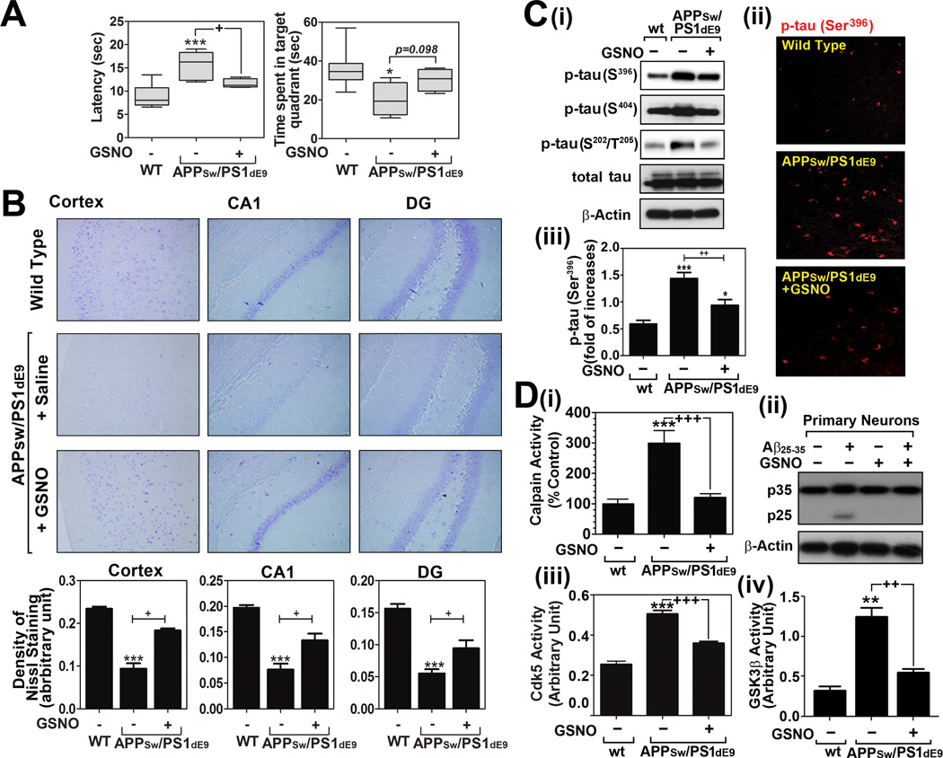
Fig. 4. Effect of GSNO treatment on spatial learning and memory deficits and tau hyperphosphorylation in of APPSw/PS1dE9 mice.
A. Analysis of cognitive function (learning and memory function) of wild type (WT/C57BL6) and APPSw/PS1dE9 mice treated with saline or GSNO (3mg/kg/day). Time (sec) to reach the hidden platform (latency in seconds) for spatial learning performance and time (sec) to spend in target quadrant without platform for spatial memory performance were analyzed by Morris Water maze test (n=8). The columns show 75% of distribution; horizontal bar in each column is median; and vertical T-bars are minimum and maximum values of the data. For statistical analysis and presentation of the data, one way ANOVA test with Turkey’s multiple comparison test was performed. B. Distribution and number of neurons in cortical and hippocampal layers (dentate gyrus; DG and CA1) were analyzed by Nissl staining. C. Brain levels of hyper-phosphorylated tau protein in WT and APPSw/PS1dE9 mice treated with saline or GSNO were analyzed by Western analysis (i) and immunofluorescence staining (ii and iii). D. Calpain activity (i), p35 proteolysis to p25 (ii), Cdk5 activity (iii), and GSK-3β activity (iv) were analyzed in brains of WT and APPSw/PS1dE9 mice treated with saline or GSNO were analyzed as described materials and methods. The vertical columns in panels B~D are means of individual data and T-bars are standard error mean. *p<0.01, **p<0.005, and ***p<0.0001 as compared to the wild type (WT) group. +p<0.01, ++p<0.005, and +++p<0.0001 as compared to untreated APPSw/PS1dE9 mice group.

