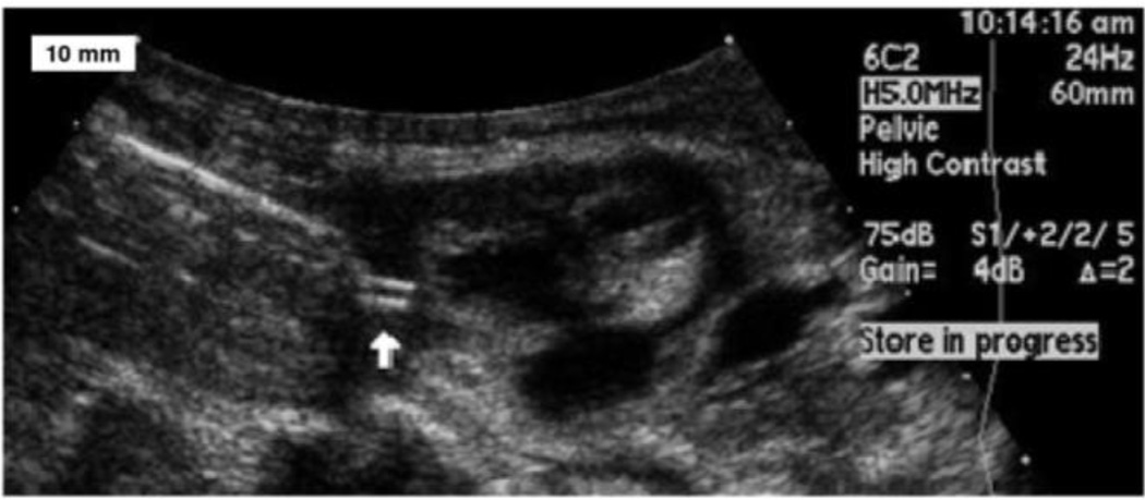
Figure 2.
An ultrasound image shows the implantation of the helical electrode (white arrow) into myocardium. An epoxy reinforcing disk between the corkscrew electrode and the helical flexible lead is wedged into the end of thin wall plastic sheath. Under trans-uterine ultrasound guidance, the trocar and cannula are advanced until it abuts the fetal heart. The trocar is then removed and the plastic sheath assembly is inserted in its place. The fetal surgeon screws the electrode into the myocardium by rotating the sheath.

