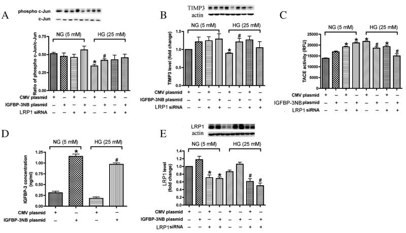
Fig. 1.
IGFBP-3 overexpression inhibited pro-inflammation markers in REC in high ambient glucose. In all experiments, REC cells were treated with IGFBP-3 plasmid and LRP1 siRNA in medium containing normal glucose (NG-5 mM) or high glucose (HG-25 mM) medium. A. Levels of IGFBP-3; B. Western Blot result of ratio of phosphor-cJun/cJun; C. Western blot result of TIMP-3 expression; D. TACE activity; E. Expression of LRP1. *P < 0.05 vs. NG control plasmid DNA transfection. #P < 0.05 vs. HG control plasmid DNA transfection. N = 3.

