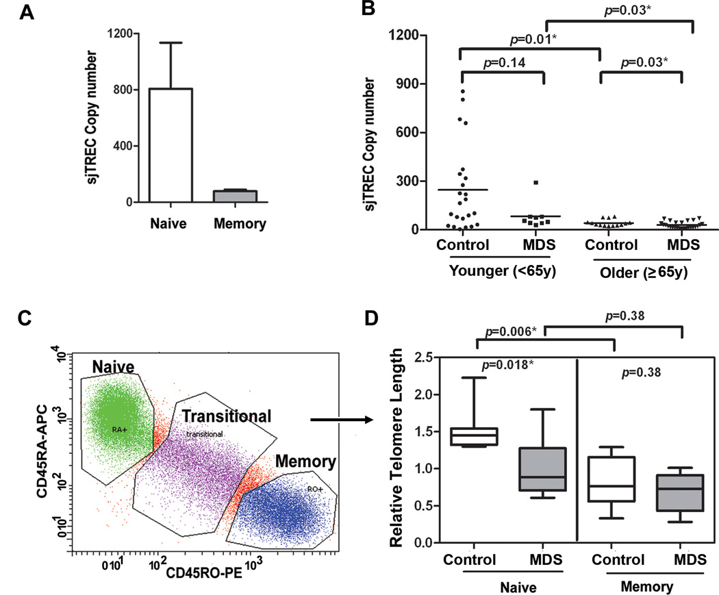
Figure 3. Telomere attrition within naïve T-cells.
(A) Naïve T-cells exclusively express sjTRECs and our data on sorted cell populations confirm that sjTREC DNA is present within the phenotype of naïve (CD45RA+CD45RO−) compared to memory (CD45RO+CD45RA−) T-cells. Using sjTREC expression as an indicator of the number of naïve cells in peripheral blood T-cells, bulk CD3+T-cells from blood of MDS patients were examined. Case-control difference in TRECs were analyzed in groups based on age<65y and ≥65y. The same DNA was used to determine telomere length in Fig 1 and at Day 0 for PD. sjTREC in peripheral CD3+ T-cell was detected using quantitative PCR and GAPDH was used as normalization for cellular DNA content in the sample. (C) Naïve and memory T-cells were isolated by flow sorting to 99% purity using the gating strategy shown. Flow cytometry was conducted after gating on viable cells and gating on CD3+ T-cells. CD45RA-APC and CD45RO-PE were used for the analysis. (D) From MDS patients (n=8, gray box) and age-matched healthy controls (n=8, white box), DNA was extracted for telomere length analysis from sorted (highly purified) naïve and memory populations. Telomere length was detected using qRT-PCR, with 293 T-cell as a calibrator, and the data was analyzed using ∆∆Ct, as described in supplemental methods. Case-control differences were compared using the Wilcoxon signed rank and p-values for the case-control differences are shown at the top of each panel. All tests were two-sided and associations were considered statistically significant at a significance level of p<0.05.

