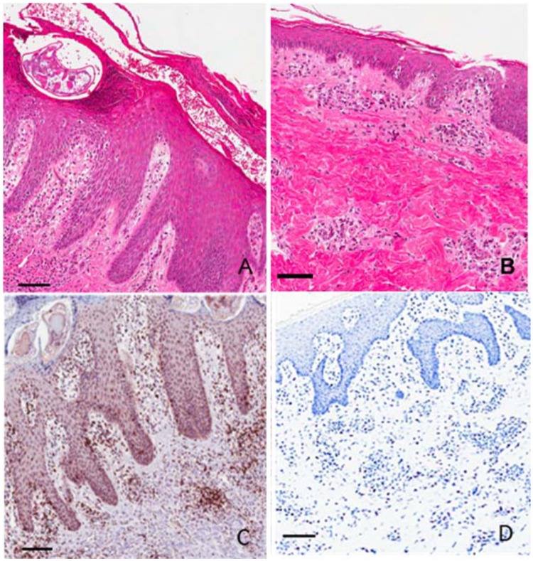
Fig 2. Representative histology and immunohistochemistry of skin lesions at week 12 post infestation with Sarcoptes scabiei.
(A) Crusted scabies, hematoxylin and eosin stain; (B) Ordinary scabies, hematoxylin and eosin stain; (C) Crusted scabies, anti-CD3+ antibody T cell stain; (D) Crusted scabies, toluidine blue mast cell stain. Scale bars = 100μM.

