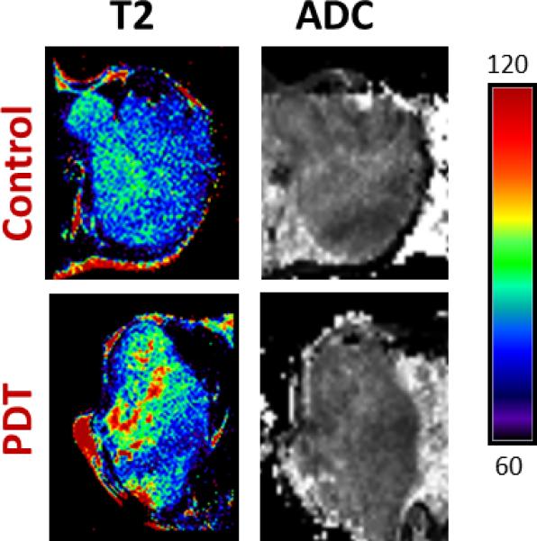
Figure 4. MR mapping of SCCVII response to PDT (transcutaneous illumination).

Panel of images represent T2 and ADC maps of a control SCCVII tumor and a tumor-treated with transcutaneous PDT. T2 mapping revealed minimal changes within the tumor following transcutaneous illumination at 24 hours post treatment. No visual evidence of photodynamic damage was seen on ADC maps following transcutaneous PDT compared to untreated control tumors.
