
FIG. 4.
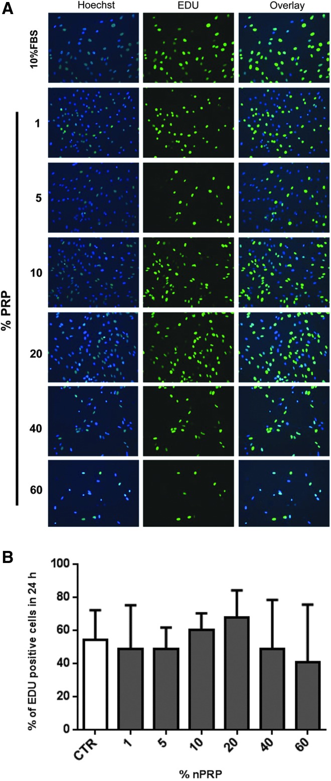
Assessment of 24 h-proliferation efficiency of AT-MSCs by EdU staining. Different concentrations of nPRP (1–60%) were compared to 10% FBS on AT-MSCs (P1) at the end of 10 days culture period. (A) Active proliferating cells are revealed by EdU-based green fluorescence nucleus staining, compared to total cells stained by Hoechst dye (blue nucleus staining). (B) Percentage of EdU positive cells was estimated by the formula: (green bright AT-MSC nucleus/total blue AT-MSC nucleus)×100. n=4.