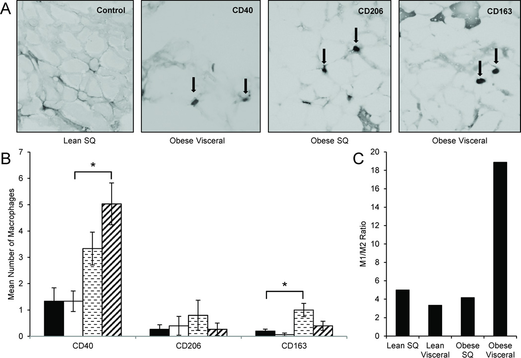
Figure 1. M1:M2 Macrophage immunohistochemistry of adipose tissues.
(A) Frozen sections of adipose from visceral and subcutaneous (SQ) depots were stained for CD40 (M1), CD206 (M2), and CD163 (M2), in 7 obese and 5 lean individuals. Representative samples are shown at 20× magnification.
(B) Obese visceral adipose shows a higher number of CD40+ macrophages than lean visceral adipose. In addition, obese subcutaneous adipose has a higher number of CD163+ macrophages than lean subcutaneous adipose. No significant differences were found for CD206+ macrophages. *P-values <0.05 determined by a two-sided Student’s t-test; error bars represent ±SEM; key: ■Lean SQ, □Lean Visceral,  Obese SQ,
  Obese Visceral.
(C) The obese visceral adipose depot showed the highest M1:M2 ratio compared to every other condition.

