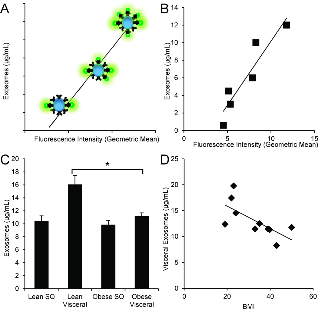
Figure 2. Quantification of adipocyte-derived exosomes using bead-based flow cytometry.
(A) Depiction of the bead-based flow cytometry technique: Increasing exosomal concentration results in increased number of exosomes attached to beads, therefore proportionally increasing geometric mean fluorescence intensity of PKH67 staining.
(B) Generated standard curve and coefficient of determination of commercially available exosomal standards (R2=0.87).
(C) Concentration of isolated exosomes from a standardized starting amount of obese (n=7) and lean (n=5) adipose. Lean visceral adipocyte-derived exosomes are significantly more concentrated than those from obese visceral adipose. *P-value<0.05 determined by a two-sided Student’s t-test; error bars represent ±SEM.
(D) As BMI increases, visceral adipocyte-derived exosome concentration decreases (R2=0.46).

