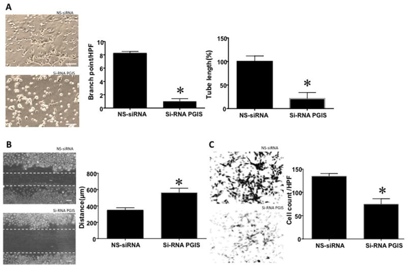
Figure 5.

A: PGIS knockdown with si-RNA decreases in vitro tube formation by control PAEC. Branch point number (n=4, p<0.05) and tube length (n=6, p<0.05) were decreased in control PAEC after application of PGIS-siRNA, compared to non-silencing (NS)-siRNA. Scale bar = 200 μ. B: PGIS si-RNA increased the gap in monolayer of control PAEC after a scratch, indicating longer recovery from injury compared to NS-RNA (n=10, p<0.05). Scale bar=100 μ. C: PGIS si-RNA also decreased the number of control cells able to invade through the Boyden chamber as compared to NS-siRNA treated control cells on cell invasion assay (n=4, p<0.05). For all studies, * indicates p<0.05 compared to NS-siRNA treated PAEC.
