
Figure 1.
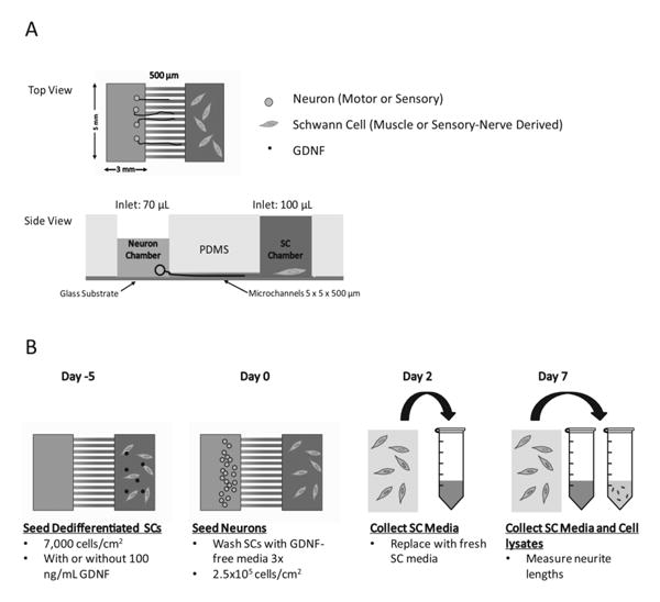
Schematic of study design and timeline for culture in microdevices. A) Microdevice and microchannels dimensions are 5 × 5 × 500 (L×W×H) μm. Volume difference allows for one way flow and diffusion of media and growth factors. B) Timeline of cell seeding, media/cell lysate collection, and neurite length analysis is shown.