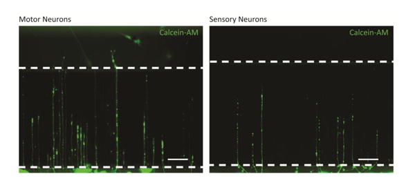
Figure 2.

Representative images of motor (A) and sensory (B) neurites extending into microchannels with support from phenotypically matched SCs in the opposing chamber. Dotted lines mark microchannel ends. Neuronal chamber, microchannels, and SC chamber are oriented from bottom to top of image. Scale bar = 100 μm.
