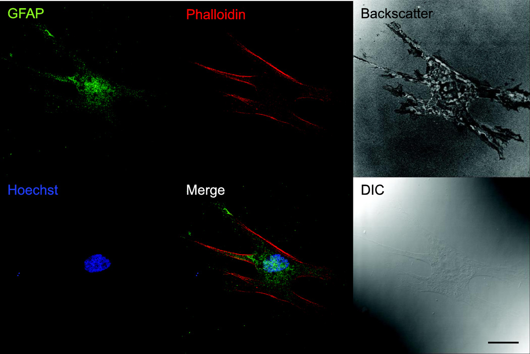
Figure 1. Cultured ONHAs are positive for GFAP.
Representative images for primary cultured rat ONHAs (passage 3) stained for GFAP, colabeled with AlexaFluor® 647 Phalloidin and Hoechst 33258. Backscatter image generated with 647 nm laser illumination shows morphology and adhesion of ONHA. A differential interference contrast (DIC) image is shown for comparison. Scale bar: 25 µm.

