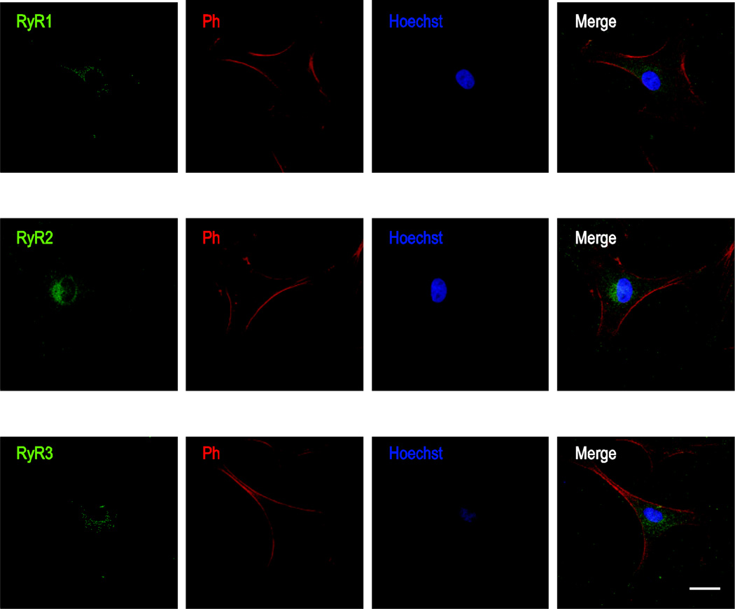
Figure 3. Immunocytochemical localization of RyRs in primary cultured rat ONHAs.
Representative images are shown for staining against RyR1 (A), RyR2 (B), and RyR3 (C), using AlexaFluor® 488-labeled secondary antibody. As for IP3R staining in Fig.1, cells were co-stained using AlexaFluor® 647 Phalloidin and Hoechst 33258. Note the punctate, cytosolic immunoreactivity indicative of ER localization typical for RyRs. We identified all RyR subtypes in primary cultured rat ONHAs, with RyR2 showing the strongest Scale bars are 25 µm.

