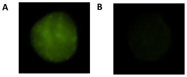
Figure 1.

Endothelial cell stained with both the primary antibody for adiponectin and the corresponding secondary antibody conjugated with the fluorescent tag (A). Negative control endothelial cell stained with the secondary antibody, but without the primary antibody for adiponectin (B).
