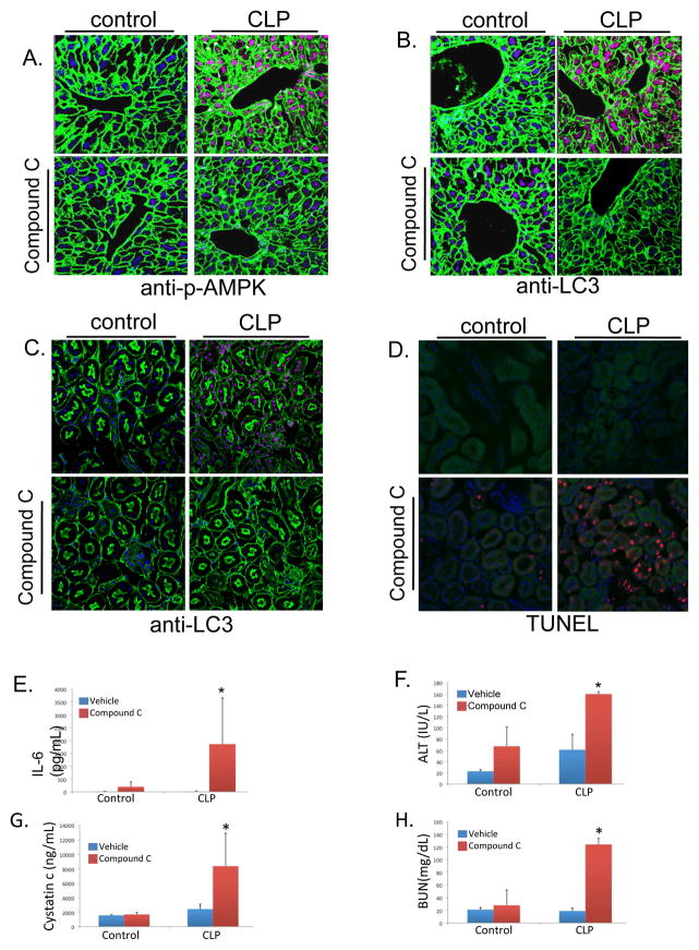
Figure 4.
Inhibition of AMPK signaling exacerbated injury and inflammation. A. CLP increased phosphorylation of AMPK as demonstrated by immunohistochemistry in liver tissue. Compound C prevented CLP-induced AMPK phosphorylation [Green=actin; blue=dapi; red=p-AMPK]. B, C. Autophagy as determined by LC3 protein levels was increased in liver (B.) and Kidney (C.) following CLP [Green=actin; blue=dapi; red=LC3]. Compound C limited these increases in LC3 levels. D. Minimal apoptotic cell death is seen following CLP, however, there is increased apoptotic cell death with inhibition of AMPK signaling as determined by TUNEL staining in kidney tissue [Green=actin; blue=dapi; red=TUNEL]. E–H. Inhibition of AMPK signaling resulted in worse inflammation and tissue injury at 24 hours. Serum IL-6 levels normalized by 24 hours following CLP, however, Compound C treated animals continued to show elevated levels at this time point (*P<0.05 compared to vehicle, CLP mice; E.). Furthermore, there was no significant difference in organ injury in vehicle treated CLP mice compared to control mice at 24 hours following CLP. Compound C pretreatment led to exacerbated injury at this timepoint (*P<0.05 compared to vehicle, CLP mice; F–H.).

