
Figure 4. Quantification of fusion rates in skeletal muscle progenitor cell populations as a function of tear state.
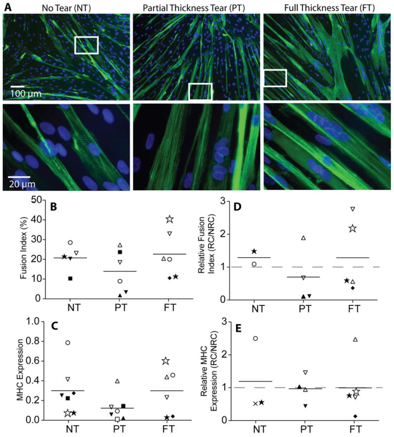
A) Immunofluorescence images of myotubes formed from the fusion of SMPs identified by myosin heavy chain (MHC, green). Striations of MHC are visible in higher magnification images. B) Quantification of the fusion index (% nuclei in myotubes) as a function of tear state. C) MHC gene expression quantified by qPCR as a function of tear state. D) Fusion index from rotator cuff (RC) muscles normalized to a patient-matched non-rotator cuff (NRC) Deltoid control. E) MHC expression from rotator cuff (RC) muscles normalized to a patient-matched non-rotator cuff (NRC) deltoid control.