
Figure 5. Engraftment of rotator cuff muscle progenitor cells into regenerating mouse muscle.
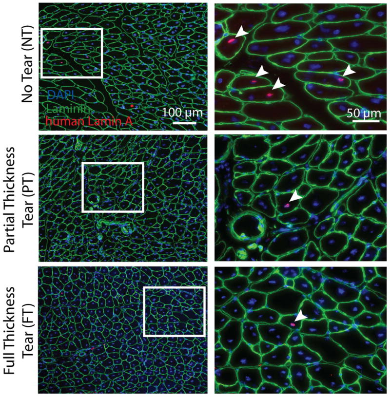
Images in the left-hand column show histological sections based on tear state. Nuclei derived from human SMPs are indentified by a human-specific lamin A antibody (red). Counterstaining with laminin (green) and DAPI (blue) demonstrate human nuclei centrally located in muscle fibers indicating that they were incorporated during regeneration (arrows). White box (left) indicates where images were magnified in right-hand column.