Figure 2.

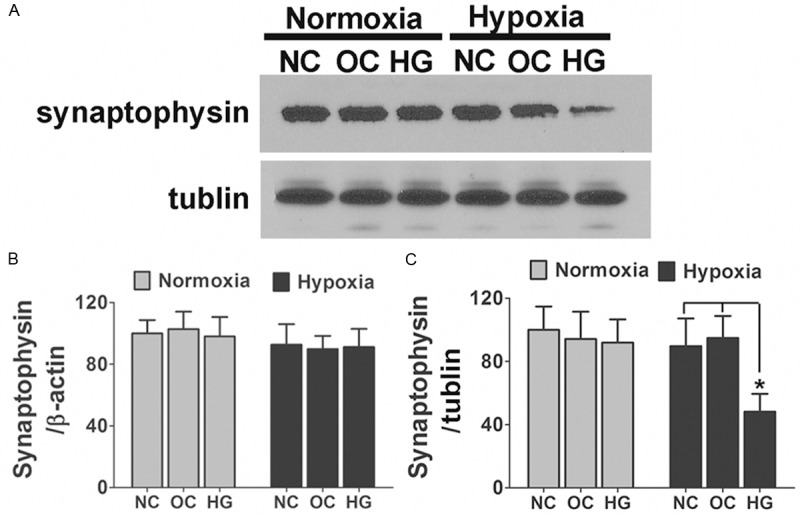
Posttranscriptional reduction of synaptophysin was detected in primary cultured hippocampus neurons after hypoxia and high glucose exposure. A. Primary cultured hippocampus neurons treated either by 50 mM glucose or 25 mM glucose + 25 mM mannitol were subjected to 3 days hypoxia or normoxia exposure. The levels of synaptophysin were analyzed by immunoblot. Shown are representative blots from six independent experiments with similar results. B. Neurons were treated as described above; the mRNA expression levels of synaptophysin are shown. There was no significant difference between different groups. Data are mean ± SD from six independent experiments. C. Densitometric quantification of synaptophysin in hippocampus neuronal cells treated with different culture medium in response to hypoxia or normoxia exposure. Data are mean ± SD from six independent experiments. *, P<0.05 vs. NC or OC group. NC, normal control; OC, osmotic control; HG, high glucose.
