
FIG 1.
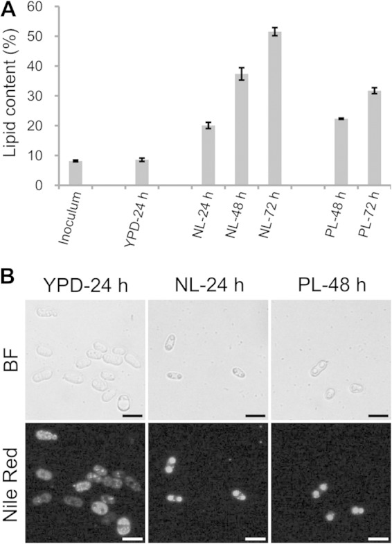
Lipid accumulation by R. toruloides cells. (A) Lipid contents of R. toruloides cells cultured under different conditions. Cells were collected from cultures in YPD, nitrogen-limited (NL), and phosphorus-limited (PL) media. The total lipids were extracted and measured gravimetrically. The error bars represent the standard deviations for three independent samples. (B) Morphology of R. toruloides lipid droplets (LDs). Cells cultured in YPD medium for 24 h, NL medium for 24 h, or PL medium for 48 h were stained with Nile Red and viewed by fluorescence microscope. BF, bright field microscopy; bar, 10 μm.