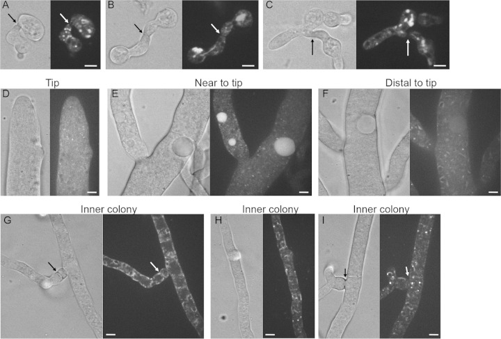
FIG 6.
Localization of LDF-2-GFP in germlings and in hyphae. (A to C) LFD-2-GFP localization in germlings undergoing chemotropic interactions and cell fusion. Arrows indicate the point at which germlings are adhered. (D to F) Cellular localization of LFD-2-GFP in apical hyphae and in hyphae more distant from the colony periphery. Note localization to punctae in apical hyphae and hyphae near the tips (D and E) but localization to nuclear ER and membrane structures in hyphae further from the periphery of a colony (F). (G to I) Localization of LFD-2-GFP to fusion hyphae, as well as hyphae within the interior of a colony. Arrows indicate the point of contact between fusion hyphae. Note the localization of LFD-2-GFP to nuclear ER, puncta, and cortical ER or plasma membrane in these hyphae. Scale bars, 5 μm.

