
FIG 2.
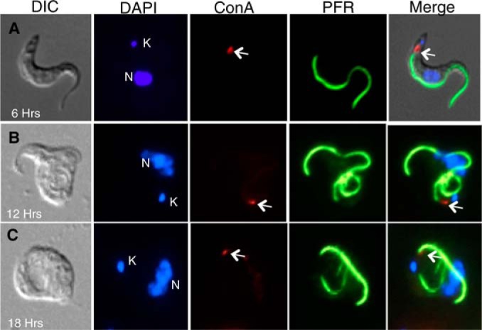
Localization of the α-KDE1 RNAi-induced vacuole. (A to C) Following induction with doxycycline for 6, 12, or 18 h, α-KDE1 RNAi T. brucei cells were incubated at 3°C with ConA-FITC, fixed, incubated with antibodies against the PFR protein, and stained with DAPI. The positions of the DAPI-stained kinetoplast (K) and nucleus (N) are indicated, as is that of bound ConA (arrow).