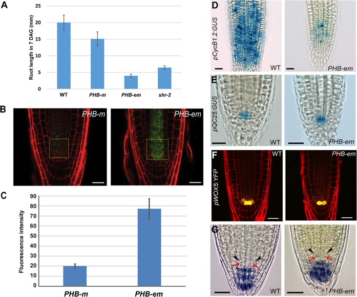
Fig 3. PHB in the stele regulates root meristem and growth activity in a QC-independent manner.
(A) A comparison of root lengths in wild-type, pWOL:PHB-m:GFPNLS, pWOL:PHB-em:GFPNLS and shr-2 plants (7 DAG). The error bars represent the standard error (n = 9–14 plants). (B, C) Quantitative comparison of PHB-GFP levels in the root stele cells expressing pWOL:PHB-m:GFP NLS and pWOL:PHB-em:GFP NLS. Fluorescence intensity was measured for GFP in the boxed area of the panel (B). Error bars represent standard error (n = 11) (D) pCycB1.2:GUS expression shows drastic reduction in cell division potential in the proximal meristem. Expression of pQC25:GUS (E) and pWOX5:YFP (F) in the PHB-em roots. (G) Starch granule accumulation as visualized by Lugol’s staining in the pWOL:PHB-em:GFP NLS roots. The black arrowheads and red arrows indicate the QC and columella stem cells, respectively. Scale bars: B, 20 μm; D-G, 25 μm.

