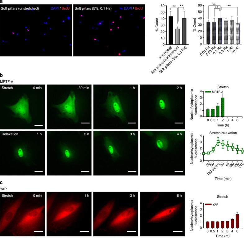
Figure 4. Effect of cyclic stretching on cell proliferation.
(a) Percentage of cells costained with 5-bromo-2′-deoxyuridine (BrdU) and DAPI on different stretching types and frequencies: unstretched flat PDMS (n=140 cells), unstretched soft pillars (n=142), 5% cyclic stretched soft pillars at 0.1 Hz (n=74), 0.01 Hz (n=93), 0.03 Hz (n=66), 0.3 Hz (n=106), 1 Hz (n=59) and 10 Hz (n=79). Error bars; s.e.m. **P<0.01; Student’s t-test. Time-dependent translocation of (b) GFP–MRTF-A (green fluorescent protein tagged Myocardin-related transcription factor-A) and (c) YAP–GFP (green fluorescent protein tagged Yes-associated protein) by cyclic stretch (5%, 0.1 Hz). Plasmids were transient transfected into PMEFs through electroporation at least 24 h before imaging. Stretching was applied 2 h after the seeding of cells in the chamber. Over 20 cells were analysed for each protein. Experiments were repeated at least three times and showed similar results. Scale bar=20 μm.

