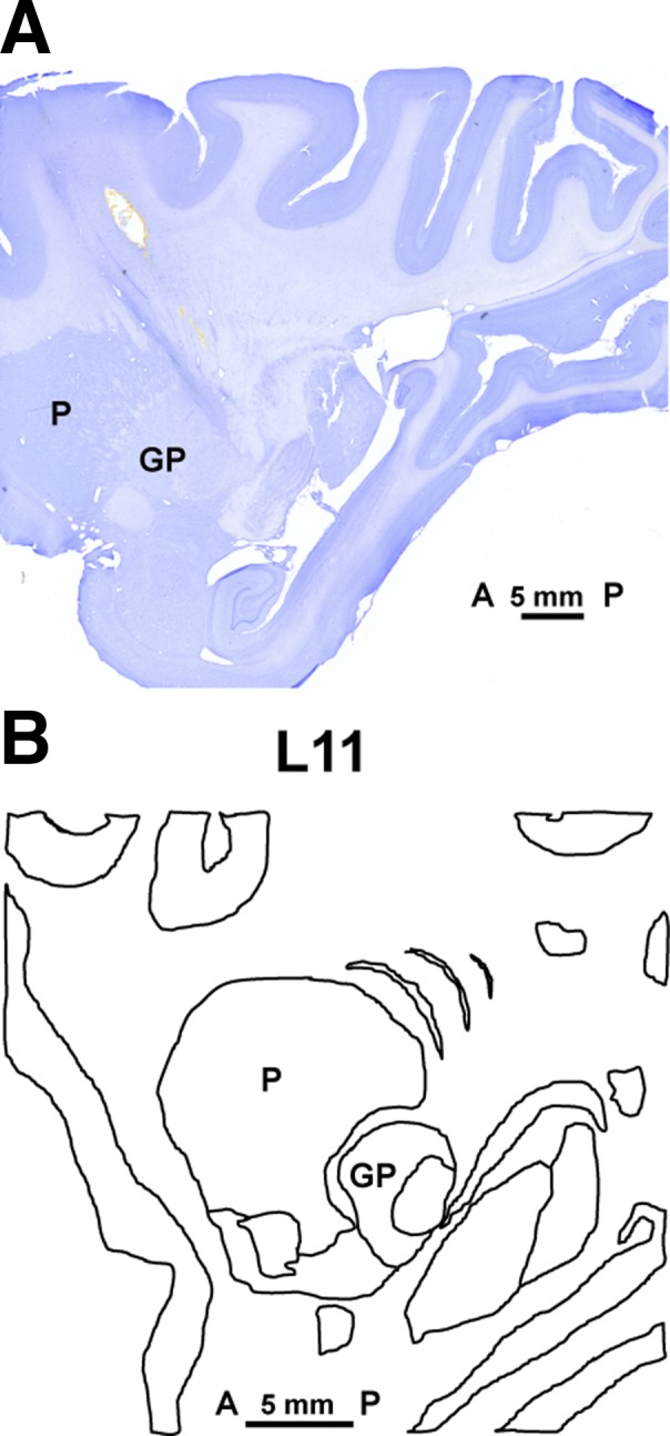
Fig. 1.

A: the Nissl-stained section of monkey brain shows the electrode track of a brain penetration made before euthanasia to show one of the recording sites. The electrode penetration targeted the posterior and lateral area of the putamen, passing through its edge according to coordinates taken from our electrophysiological mapping. The section matched the anatomy of lateral 11 (L11) in the sagittal plane of the monkey atlas (B). To confirm the location of recorded units, the anatomic position of electrode penetrations used in the recordings was then calculated with reference to the coordinates of this electrode track in L11. Bar shows the scale from anterior (A) to posterior (P). L, lateral; P, putamen; GP, globus pallidus.
