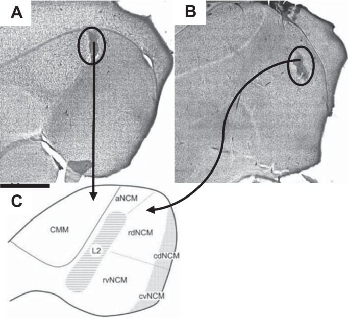
Fig. 1.

Histology and electrode placement in caudomedial nidopallium (NCM) and caudomedial mesopallium (CMM). Sites used for data analyses were confirmed to be in either CMM (A) or NCM (B) by sending electrolytic current (20 μA for 12 s) through electrodes at the conclusion of the electrophysiological experiment to produce lesions (scale bar = 500 μm). Brains were then sectioned, and slices were stained with cresyl violet and visualized under a light microscope to confirm placement. C: figure showing the boundaries of avian auditory areas (NCM, L2, CMM). aNCM, rdNCM, rvNCM, cdNCM, and cvNCM: apical, rostrodorsal, rostroventral, caudodorsal, and caudoventral NCM, respectively. [From Sanford et al. (2010). Reprinted with permission from Wiley.]
