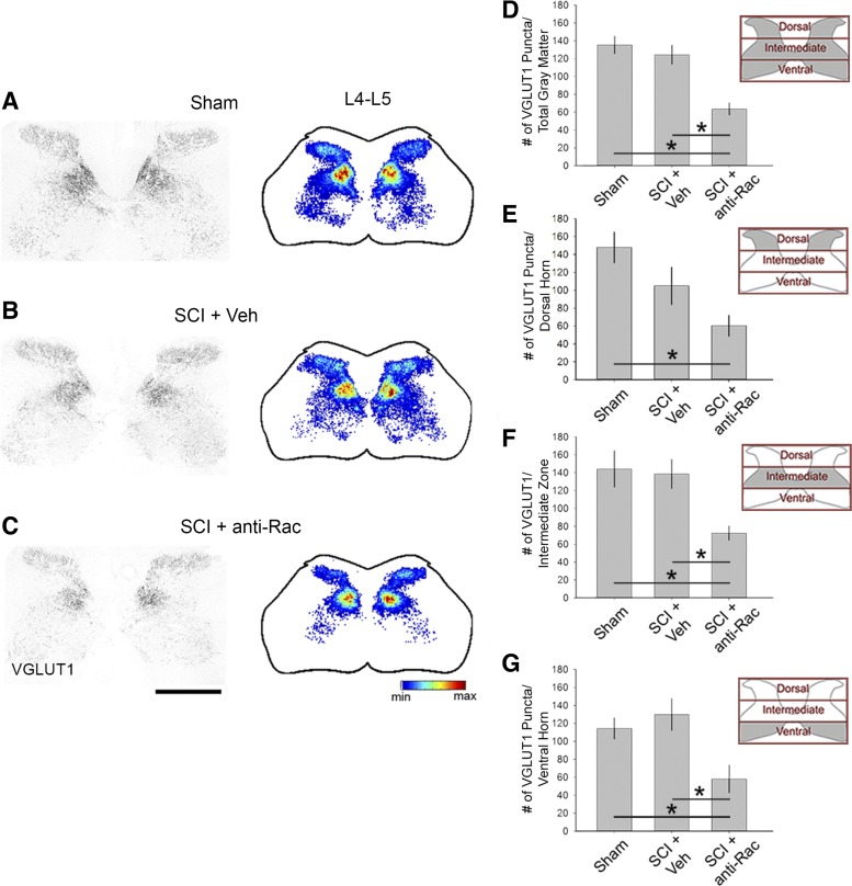
Fig. 9.
Excitatory terminals in the spinal cord gray matter. Vesicular glutamate transporter 1 (VGluT1)-immunopositive puncta appeared throughout all laminae of the spinal cord gray matter in the lumbar enlargement L4–L5 (A–C, left). Spatial heat maps (A–C, right; red = highest density, blue = lowest density) shows the overall areal density of VGluT1 expression in sham (A), SCI + Veh (B), and SCI + anti-Rac (C) treatment groups. Quantification of the VGluT1 puncta within the total gray matter region (D), dorsal horn (E), intermediate zone (F), and ventral horn (G) as represented in insets as gray shading demonstrates no significant change in SCI + Veh compared with sham. Treatment with the Rac1 inhibitor in SCI animals decreased VGluT1 areal density compared with SCI + Veh in the total gray matter, intermediate zone, and ventral horn only (*P < 0.05) with no significant change in the dorsal horn (P > 0.05). The areal density of VGluT1 decreased in SCI + anti-Rac1 compared with sham in the dorsal horn (*P < 0.05). Scale bar for A–C, 500 μm.

