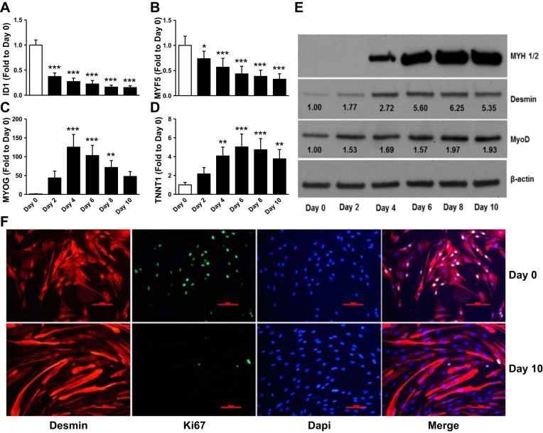
Fig. 1.
Validation of myogenic marker expression during human skeletal muscle cell differentiation. Skeletal muscle cells cultured in vitro as myoblasts and induced to differentiate were harvested at various time points during differentiation for analysis of gene expression (A–D), protein abundance (E), or immunocytochemistry (F). A–D: quantitative reverse transcriptase-polymerase chain reaction (qPCR) analysis of proliferative and myogenic markers. White bars, proliferative cells (day 0); black bars, different days following differentiation induction. E: Western blot analysis of myogenic markers. When appropriate numbers inserted on the immunoblot indicate average fold change over day 0. Representative blots. F: immunocytochemistry the myogenic marker desmin, proliferative marker Ki67, and DAPI of skeletal muscle cells before and after 10 days of differentiation. Scale bars are 100 μm. Representative images (n = 3). Data are means ± SE across biological replicates (n = 6). *P < 0.05, **P < 0.01, ***P < 0.001 by repeated-measures ANOVA.

