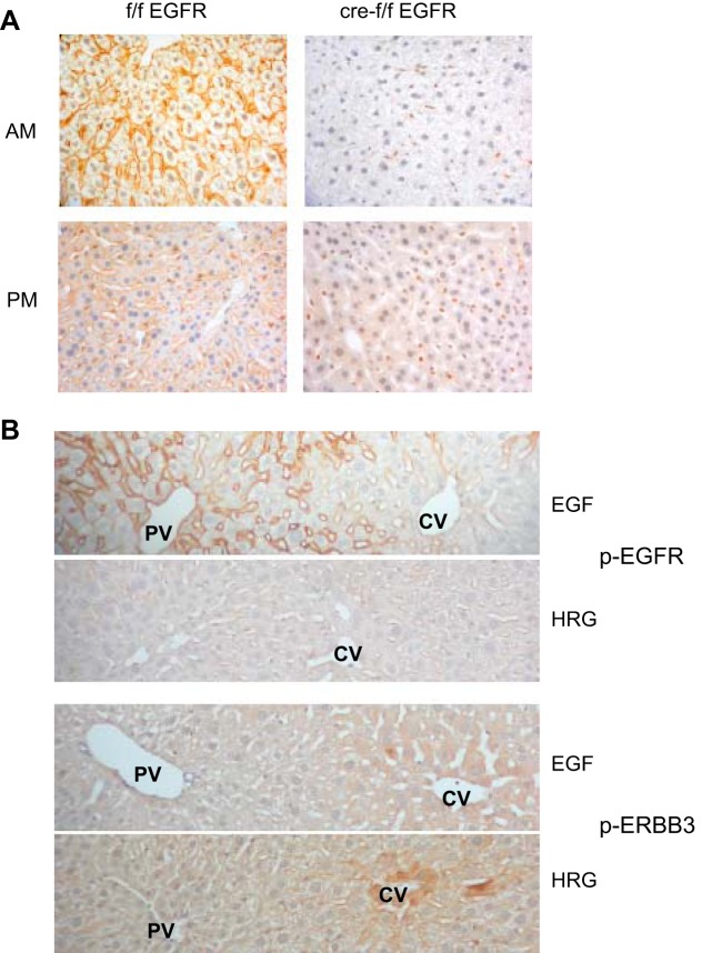
Fig. 2.
Immunohistochemical localization of EGFR and phospho-EGFR and phospho-ERBB3. A: we evaluated the immunohistochemical localization of EGFR in the liver in wild-type (left) and HS-EGFRKO mice (right) 2 h after lights on (AM) and 2 h after lights off (PM). Only a few hepatocytes expressed EGFR in the HS-EFGRKO mice. Nonparenchymal cells continued to express EGFR in the HS-EGFRKO livers. The expression in the early light phase was stronger than that in the early dark phase. Hepatocytes at the beginning of the light phase are larger because of glycogen synthesis during the nighttime feeding phase. B: we injected EGF or HRG into the portal vein (PV) and then used immunohistochemistry to identify the localization of the phosphorylated forms of EGFR (top 2 panels) or ERBB3 (bottom 2 panels). Clear differences in zonal distribution of the 2 receptor tyrosine kinases were observed. The phosphorylated form of EGFR had a striking periportal localization (in contrast to the relatively uniform distribution of “total” EGFR as shown in Fig. 2A) whereas phosphorylated ERBB3 tended to be stronger around the central vein (CV).

