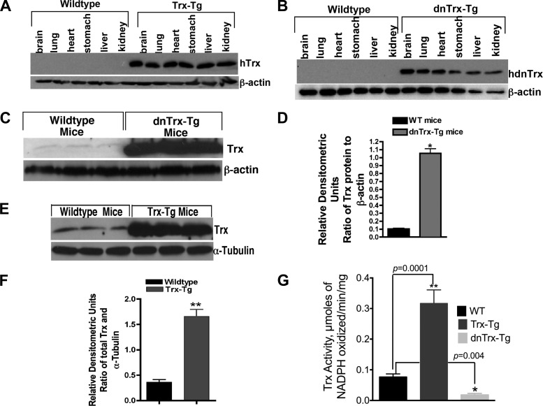
Fig. 1.
Mice with decreased levels of functional thioredoxin (dnTrx-Tg) and overexpressing Trx (Trx-Tg) show increased levels of immunoreactive Trx in various organs including the lung. F1 mice were killed, and protein expression was analyzed in various organs by immunoblot analysis using an anti-Trx antibody reactive to both human and mouse antigen. Blots were reprobed for β-actin expression to confirm equivalent protein loading. A: Trx-Tg mice. B: dnTrx-Tg mice. C: expression of Trx in the lungs of wild-type (WT) or Trx-Tg mice. D: densitometry of Fig 1C; *P < 0.05, Student's t-test. E: expression of Trx in the lungs of WT or dnTrx-Tg mice. F: densitometry of Fig 1E; **P < 0. 05, Student's t-test. G: Trx activity in the lungs of WT, Trx-Tg, and dnTrx-Tg mice; *P = 0.04; **P = 0.0001.

