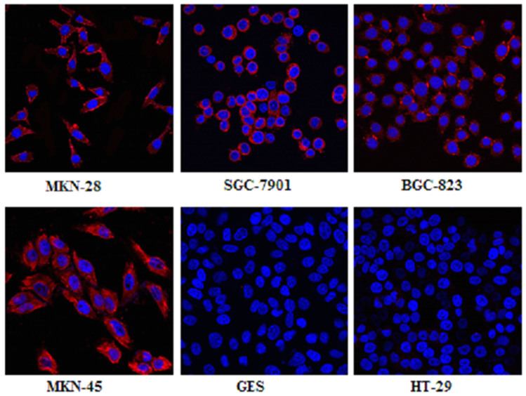
Figure 2. Laser scanning confocal microscopy imaging of different cell lines (MKN-28, SGC-7901, BGC-823, MKN-45, and GES, HT-29) after incubation with MG7 antibody showed that the expression of MG7-Ag was in the cell membrane and cytoplasm in each of gastric cell line, and there was little MG7-Ag expression in the normal gastric epithelial cell lines (GES) and colon cancer cell lines (HT-29).
All images were acquired under the same condition and displayed at the same scale. Magnification: 60×.

