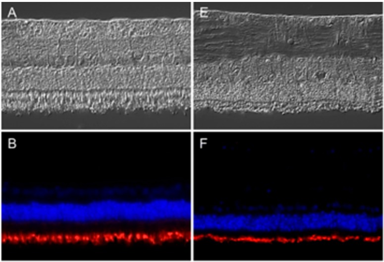
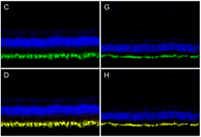
Figure 1.
Expression pattern of peripherin 2 (red) and rhodopsin (green) in representative retinas of healthy (left) and equine recurrent uveitis (ERU) cases (right). Differential interference contrast image of healthy (A) and diseased (E) equine retina. Peripherin 2 (red) was equally distributed in the photoreceptor outer segments of healthy (B) and diseased (F) equine retina. Cell nuclei were stained with 4',6-diamidino-2-phenylindol (DAPI) (blue); Rhodopsin (green) was also equally distributed in photoreceptor outer segments of healthy (C) and uveitic (G) retinas. The overlap of peripherin 2 and rhodopsin in healthy (D) and uveitic (H) retina show overlapping expression pattern of both proteins (yellow).


