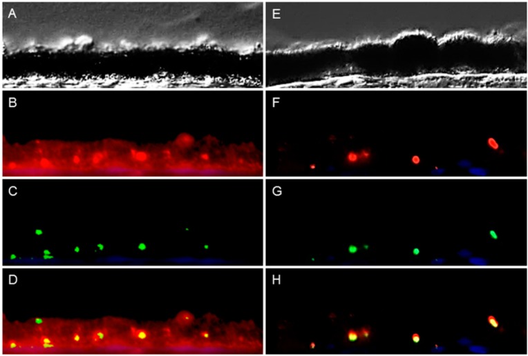
Figure 2.
Peripherin 2 (red) and rhodopsin (green) expression in retinal pigment epithelium (RPE) of representative healthy (left) and ERU cases (right). Differential interference contrast image of healthy (A) and diseased (E) equine RPE. In healthy (B) RPE, peripherin 2 was expressed over the entire RPE cell layer, and the intensity was enhanced in some spots. In uveitic RPE cells (F), peripherin 2 was significantly reduced to few small spots; Rhodopsin was limited to few isolated spots, distributed throughout the whole RPE layer, in healthy (C), as well as in diseased (G) RPE. The overlap of peripherin 2 and rhodopsin in healthy (D) RPE indicates significant higher expression of peripherin 2 compared to rhodopsin (the yellow overlap is only displayed in a few spots). In uveitic RPE (H), peripherin 2 was only expressed at rhodopsin expression sites (yellow spots).

