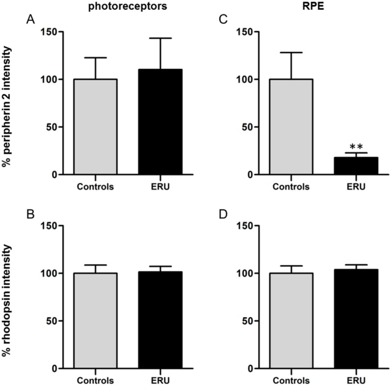
Figure 3.
(A) Quantification of the fluorescence intensity of peripherin 2 expression in photoreceptor outer segments revealed no significant difference between healthy (grey column) and ERU cases (black column); (B) There was also no significant difference between the fluorescence intensity of rhodopsin in photoreceptor outer segments of healthy (grey column) and uveitic (black column) retinas detectable; (C) A significant reduction of the fluorescence intensity of peripherin 2 in uveitic RPE to 17.9% (black column), compared to RPE of negative controls (** p ≤ 0.01); (D) Quantification of the fluorescence intensity of rhodopsin in RPE showed no significant difference between healthy (grey column) and ERU cases (black column).

