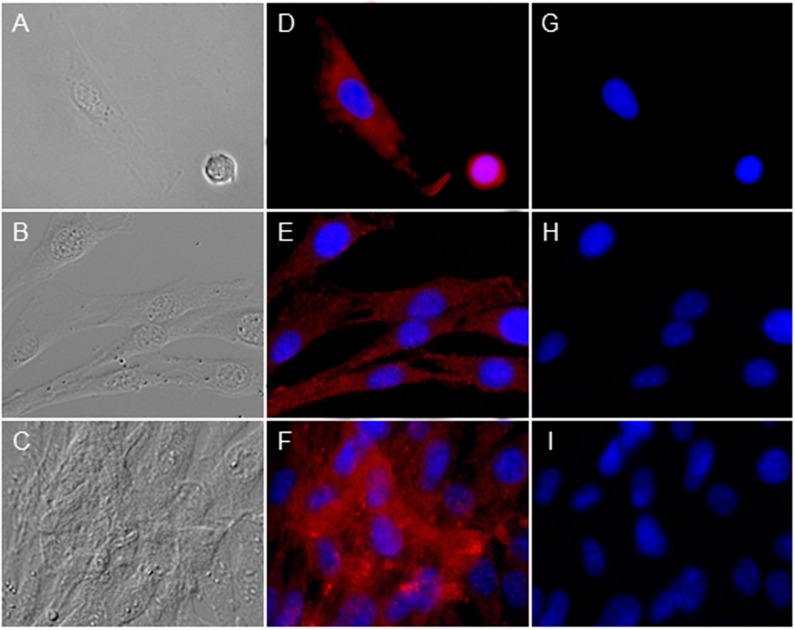
Figure 4.
Investigation of peripherin 2 (red) and rhodopsin (green) expression on cultivated healthy RPE cells at different confluency states. Differential interference contrast image of healthy equine RPE cells at different confluency states; from single cells (A) to 40% confluent cells (B) up to 90%–100% confluent cells (C); (D–F) Peripherin 2 (red) was expressed over whole RPE cells with an even intensity (cell nuclei = blue); (G–I) No rhodopsin (green) expression was found in primary healthy equine RPE cells (cell nuclei = blue).

