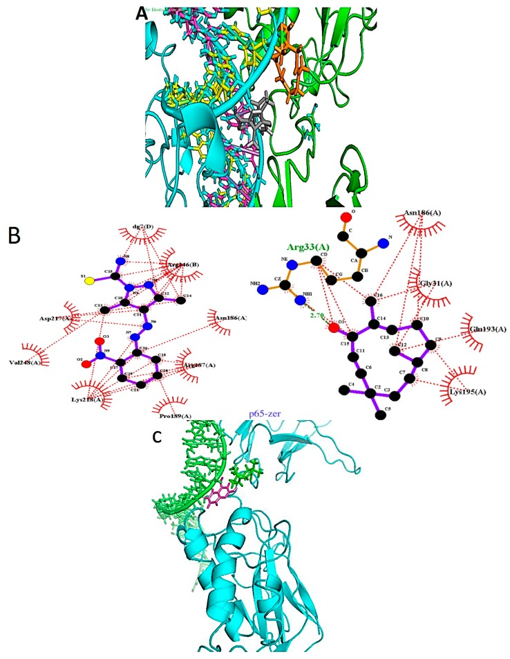
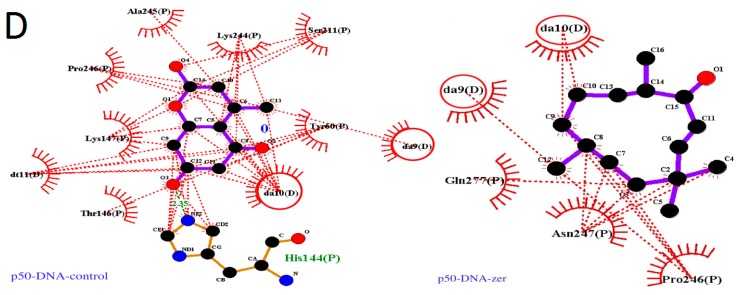
Figure 4.
Binding of reported ligands and zerumbone to nuclear factor κB. (A) p65 component: Zerumbone (orange) is placed between the chain A (green) and the DNA ladder. The known ligand (grey) can be placed between the chain A and the major groove of the DNA. Chain B of the p65 is colored cyan; (B) The LIGPLOT image of the binding site showing interactions between known ligand and zerumbone with the pocket residues; (C) p50 component: Zerumbone (green) is placed between the p50 chain (cyan) and the DNA ladder (green). The known ligand (magenta) is embedded deeper in the major groove of the DNA; (D) The LIGPLOT image of the binding site showing interactions between known ligand and zerumbone with residues in the binding the pocket.


