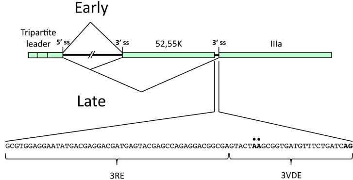
Figure 2.
Schematic drawing of the regulation of Ad5 L1 alternative RNA splicing. The early and the late expression pattern are represented in the upper and lower part of the drawing respectively. In the inset, the sequences of the IIIa repressor element (3RE) and of the IIIa virus-infection dependent splicing enhancer (3VDE) are indicated. The black dots mark the IIIa branch sites and the AG in bold the IIIa 3' splice site.

