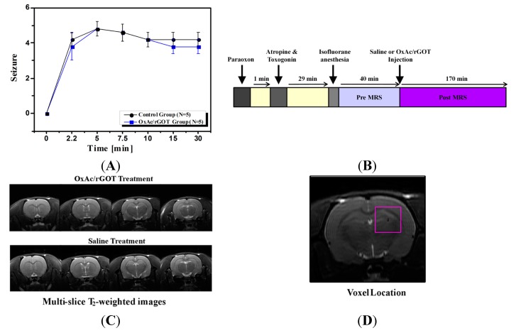
Figure 1.
(A) Racine’s scale evaluation of seizure’s post paraoxon (PO) exposure; (B) magnetic resonance spectrocopy (MRS) experiment protocol scheme; (C) four axial T2 (spin–spin relaxation)-MR-weighted images of representative rat brains treated with saline or oxaloacetate (OxAc)/glutamate-oxaloacetate transaminase (rGOT). The field of view of the the image was 4 × 4 cm2; (D) Axial MR image of the rat brain obtained by RARE (rapid acquisition with relaxation enhancement) sequence showing the position of the selected voxel (4 × 4 × 4 mm3) covering predominantly the hippocampus region. The field of view of the entire image was 4 × 4 cm2.

