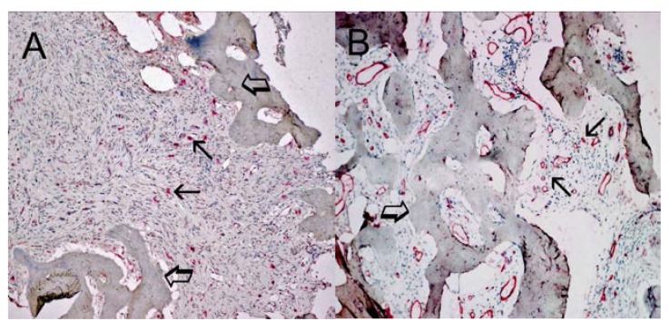
Figure 4.
(A) Bone metastasis from gastric cancer tissue section. Many scattered mast cells positive to tryptase are seen as immunostained red. Small arrows indicate single mast cells. Large arrows indicate bone tissue. Low magnification: 100×; (B) Bone metastasis from gastric cancer tissue section. Many red immunostained microvessels are present. Small arrows indicate clusters of microvessels; note the small lumen. Large arrows indicate bone tissue. Low magnification: 100×.

