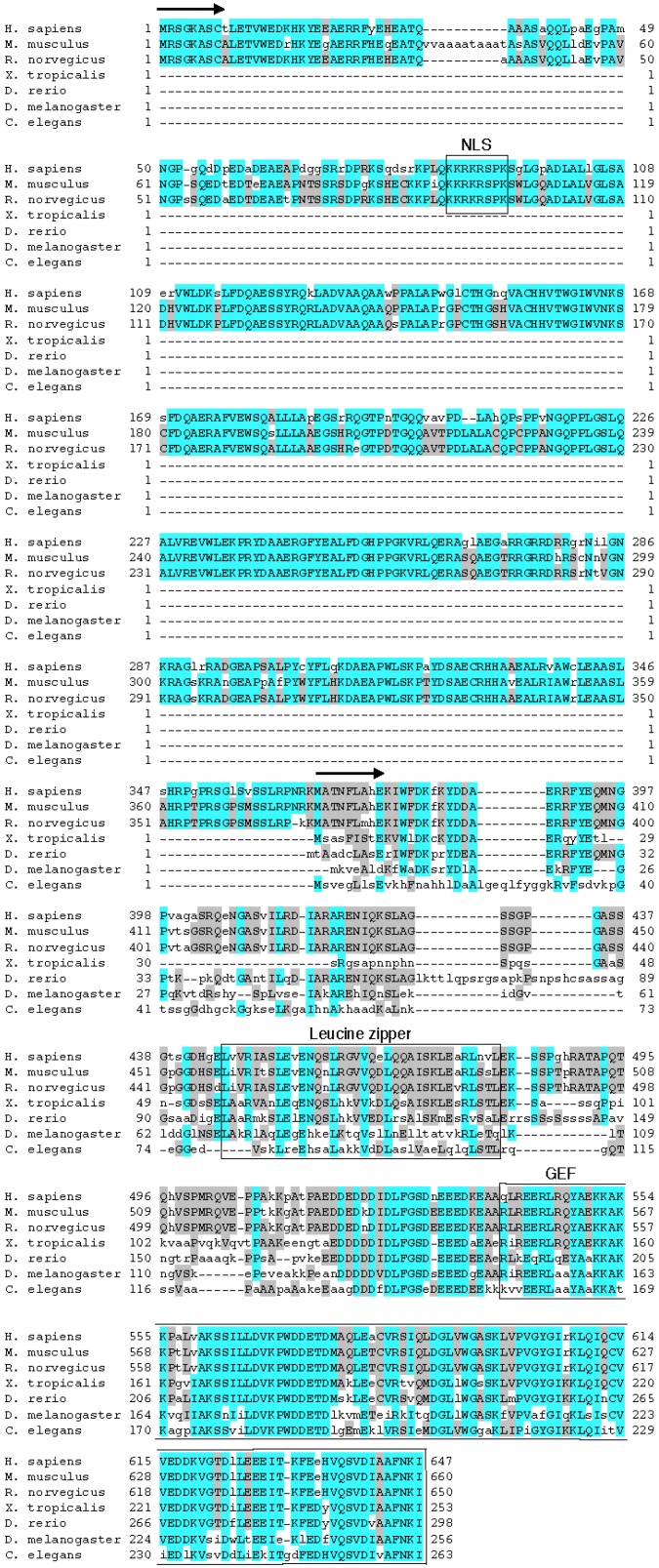
Figure 3.
Alignment of the amino acid sequences of mammalian eEF1BδL and other eukaryotic eEF1Bδ proteins. Within the compared sequences, the blue highlights show primary conserved regions, and the gray highlights show secondary conserved regions. Arrows indicate each N-terminus of the mammalian eEF1BδL and other eukaryotic eEF1Bδ proteins. The nuclear localization signal (NLS), leucine-zipper motif and guanine nucleotide exchange factor (GEF) domain are marked by boxes and text. MUSCLE was used to create the alignment [12].

