
Figure 4.
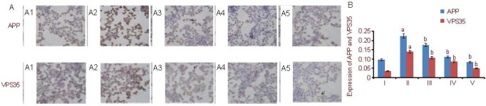
Amyloid precursor protein (APP) and vacuolar protein sorting 35 (VPS35) expression in PC12 cells (streptavidin-peroxidase method).
(A) Immunocytochemical staining of APP and VPS35 expression (endochylema staining, optical microscope, ×400).
(A1) Control group: cell nuclei are dyed blue, APP and VPS35 endochylema staining is brown (arrows).
(A2) Aβ25-35 group: endochylema staining (arrows) is much darker compared with the control group.
(A3–A5) Schisandrin B groups (5 μM, 10 μM, 25 μM): endochylema staining (arrows) gradually becomes lighter compared with the Aβ25-35 group.
In each group, endochylema staining of APP is more intense than that of VPS35.
(B) Quantification of APP and VPS35 protein expression. The data are expressed as mean ± SD and analyzed by one-way analysis of variance and least significant difference method. Image Pro Plus software was used to measure the expression of target proteins. The experiments were repeated at least three times (n = 3). aP < 0.05, vs. control group; bP < 0.05, vs. Aβ25-35 group. Furthermore, there were significant differences (P < 0.05) between the three Schisandrin B groups with different concentrations. Aβ: Amyloid β protein; I–V: control, Aβ25-35, 5, 10, 25 μM Schisandrin B groups, respectively.