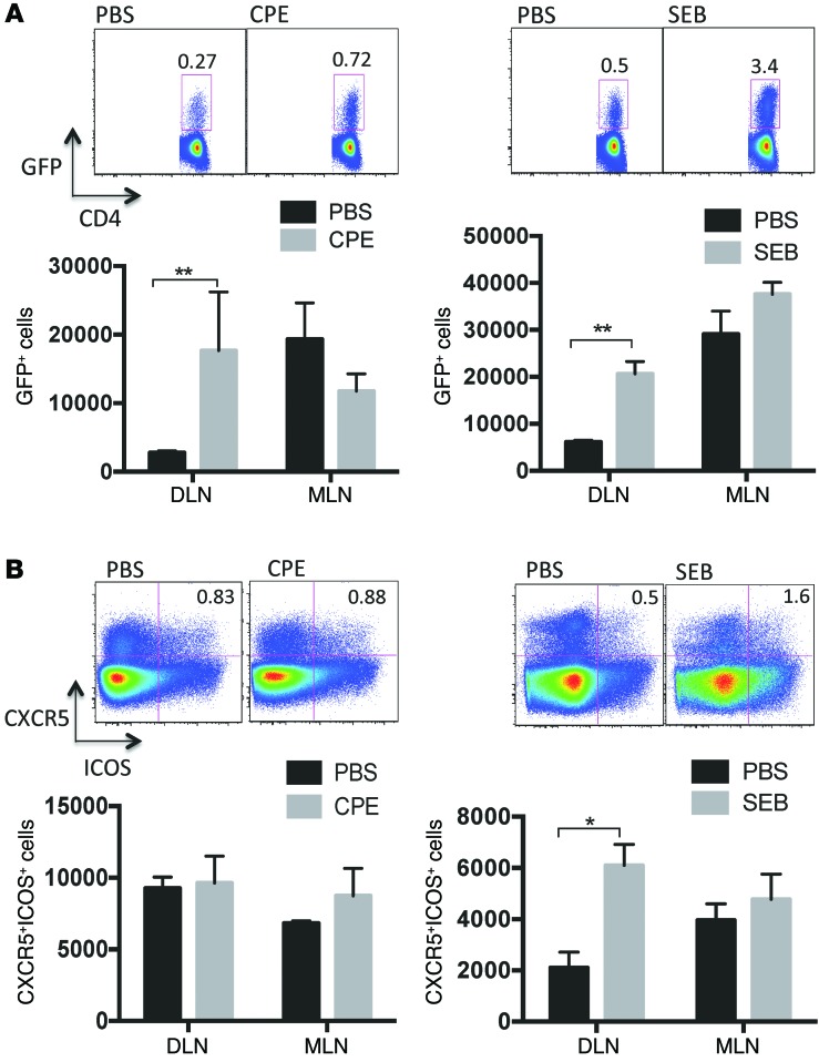
Figure 5. Effect of CPE or SEB on Th2 and Tfh cells.
4get mice were epicutaneously exposed to PBS, peanut (CPE), or SEB. One week later, skin-draining lymph nodes (DLN) and mesenteric lymph nodes (MLN) were assessed for (A) IL-4 reporter expression or (B) Tfh markers CXCR5 and ICOS. Representative plots of IL-4+ or CXCR5+ICOS+ cells of total CD4+ cells in skin-draining lymph nodes are shown above summary plots of the mean ± SEM number of cells normalized per million CD4+ T cells for 3 mice per condition. *P < 0.05, **P < 0.01.

