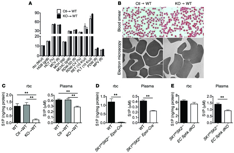
Figure 3. Adult erythropoiesis in the absence of Sphk enzymes.
(A) Lethally irradiated WT mice were reconstituted with control (Ctl) or RBC Sphk dKO (KO) fetal liver cells. Peripheral blood cell counts for the transplanted mice. HGB, hemoglobin; HCT, hematocrit; MCV, mean corpuscular volume; MCH, mean corpuscular hemoglobin; MCHC, mean corpuscular hemoglobin concentration; RDW-SD, red cell distribution width-standard deviation; RDW-CV, red cell distribution width-coefficient of variation; RET, reticulocytes; PLT, platelets; PDW, platelet distribution width; MPV, mean platelet volume. (B) Eosin staining and transmission electron microscopy images of rbc. Original magnification, ×63 and ×15,000, respectively. Plasma and rbc S1P levels in (C) transplanted mice, (D) WT and Sphk1fl/fl Sphk2+/– Epor-Cre mice, and (E) Sphk1fl/fl Sphk2–/– and EC Sphk dKO mice. n = 3 per group. **P < 0.01. All data represent the mean ± SD.

