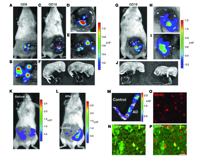
Figure 2. Development of an endometrium-specific gene delivery method: persistent transgene expression throughout pregnancy after LV-Fluc/GFP transfection of endometrial stromal cells.
(A–F) First pregnancy. Bioluminescence imaging of uteri during laparotomy on GD8 (A and B) and GD19 (C–F). There was a marked increase in Fluc signal at the implantation sites on GD8 (A and B) that was maintained through GD19 (C and D). The signal persisted in the uterine wall (E) after delivery of the fetuses and placentas (F) by cesarean section. (G–J) Second pregnancy, following mating 30 days after first delivery. As in the first pregnancy, the signal increased during pregnancy (G–I) and persisted in the uterine wall (I) after removal of the fetuses and placentas (J) by cesarean section on GD19. (K–P) Artificial decidualization. (K) Live bioluminescence imaging before induction of artificial decidualization (AD) in LV-Fluc/GFP–transfected uterus (both horns). (L) Bioluminescence imaging 3 days after artificial decidualization induction in 1 horn. Note the dramatic increase in Fluc signal in the decidualized horn (M). (N–P) Colocalization (P) of GFP (N) and BrdU (O) to decidual cells; numerous GFP-expressing cells were BrdU positive, which suggests that the increase in Fluc signal during pregnancy may have resulted from proliferation of LV-Fluc/GFP–transfected endometrial cells. Scale bar: 20 μm (N–P).

