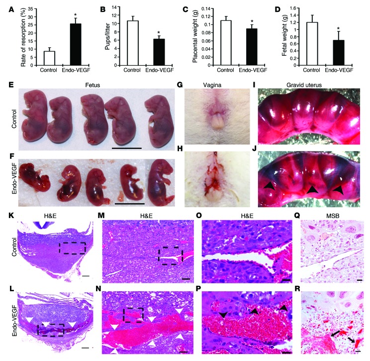
Figure 4. Overexpression of VEGF in the endometrium leads to increased pregnancy loss and widespread vascular deformity in the placenta, specifically in the venous channels and veins at the maternal-fetal interface.
(A–F) VEGF overexpression significantly increased resorption rate (A) and significantly decreased number of pups per litter (B), placental weight (C), and fetal weight (D–F) on GD18. (G and H) Vaginal bleeding occurred in most animals overexpressing VEGF, starting around GD10. (I and J) Gravid uteri in Endo-VEGF animals showed widespread hemorrhaging (arrowheads) compared with controls on GD12. (K–R) Histology of placental sections from control (K, M, O, and Q) and Endo-VEGF animals (L, N, P, and R), assessed by H&E (K–P) or martius scarlet blue (MSB) staining for extravasated fibrin (Q and R). Endo-VEGF animals showed severe dilation and congestion of venous sinuses and veins (white arrowheads) at the maternal-fetal junctions (L and N). Some of these venous sinuses exhibited focal, missing endothelial linings and hemorrhaging (black arrowheads) and extravasated fibrin (arrows) dissecting into the adjacent tissues (P and R). Results are mean ± SD. *P < 0.05 (n = 15). Scale bars: 10 mm (E and F); 500 μm (K and L); 100 μm (M and N); 20 μm (O–R).

