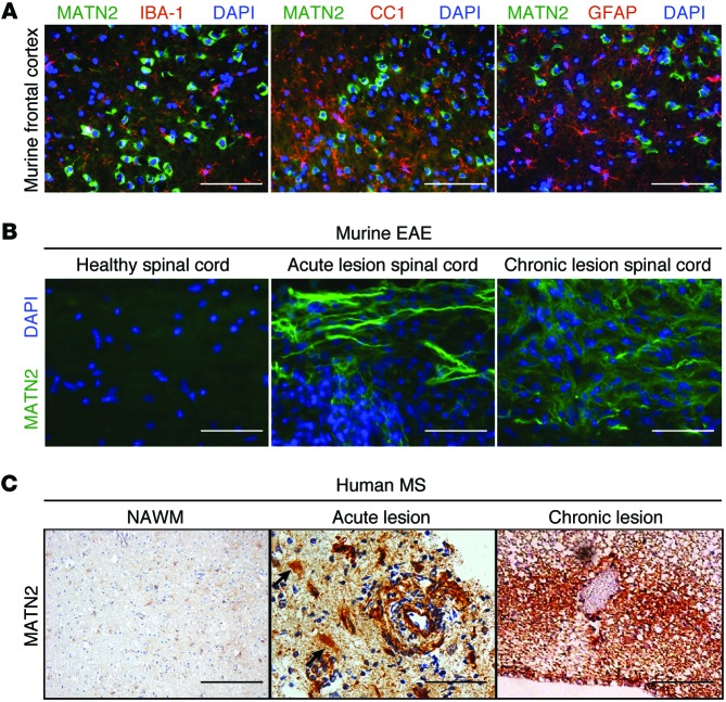
Figure 3. MATN2 expression is specific to neurons in nonlesioned EAE cortex but differentially expressed in murine EAE and human MS lesions.
(A) Colabeling for MATN2 and IBA-1, CC1, or GFAP in the frontal cortices of healthy mice. Scale bar: 50 μm. (B) Labeling for MATN2 on longitudinal spinal cord sections of healthy, acute, and chronic EAE lesions. Scale bar: 12.5 μm. (C) IHC labeling for MATN2 in normal-appearing white matter (NAWM) and early acute and chronic human MS lesions. Acute lesions show lesional extracellular, astrocytic (black arrows), and perivascular expression of MATN2. Scale bar: 200 μm (NAWM); 50 μm (acute); 100 μm (chronic).

