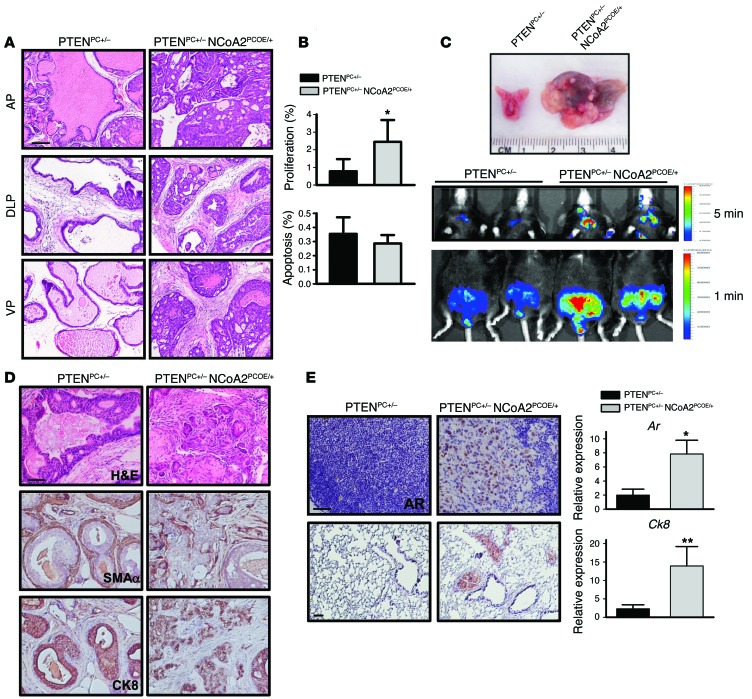
Figure 2. NCoA2 collaborates with PTEN loss to promote PCa progression and metastasis.
(A) H&E-stained sections of representative AP, DLP, and VP at 6–7 months of age in PTENPC+/– and PTENPC+/– NCoA2PCOE/+ mice. (B) The percentage of proliferating and apoptotic cells in PTENPC+/– and PTENPC+/– NCoA2PCOE/+ mice at 6 months of age were determined using Ki67 and cleaved caspase 3 assays, respectively. (C) Top: Macroscopic dissection image of prostate isolate from PTENPC+/– and PTENPC+/– NCoA2PCOE/+ mice. Representative images of luciferase-reporter activity of PTENPC+/– and PTENPC+/– NCoA2PCOE/+ mice at 12 months of age are displayed. Middle: Luciferase activity of PCa cells that have metastasized to distant sites in the chest area (exposed for 5 minutes). Bottom: Luciferase activity in the prostate and surrounding area (exposed for 1 minute). (D) H&E-stained sections of DLP at 12 months of age and IHC analyses of α-SMA and CK8 in prostate tumors. (E) Left: AR expression in lumbar lymph nodes (top) and lungs (bottom) at 12 months of age. For quantitative results of metastatic incidence, see Table 1. Right: Relative mRNA levels of Ar and Ck8 levels in lymph node from PTENPC+/– and PTENPC+/– NCoA2PCOE/+ mice (n = 6). Scale bars: 50 μm (A, D, and E). *P < 0.05; **P < 0.01.

